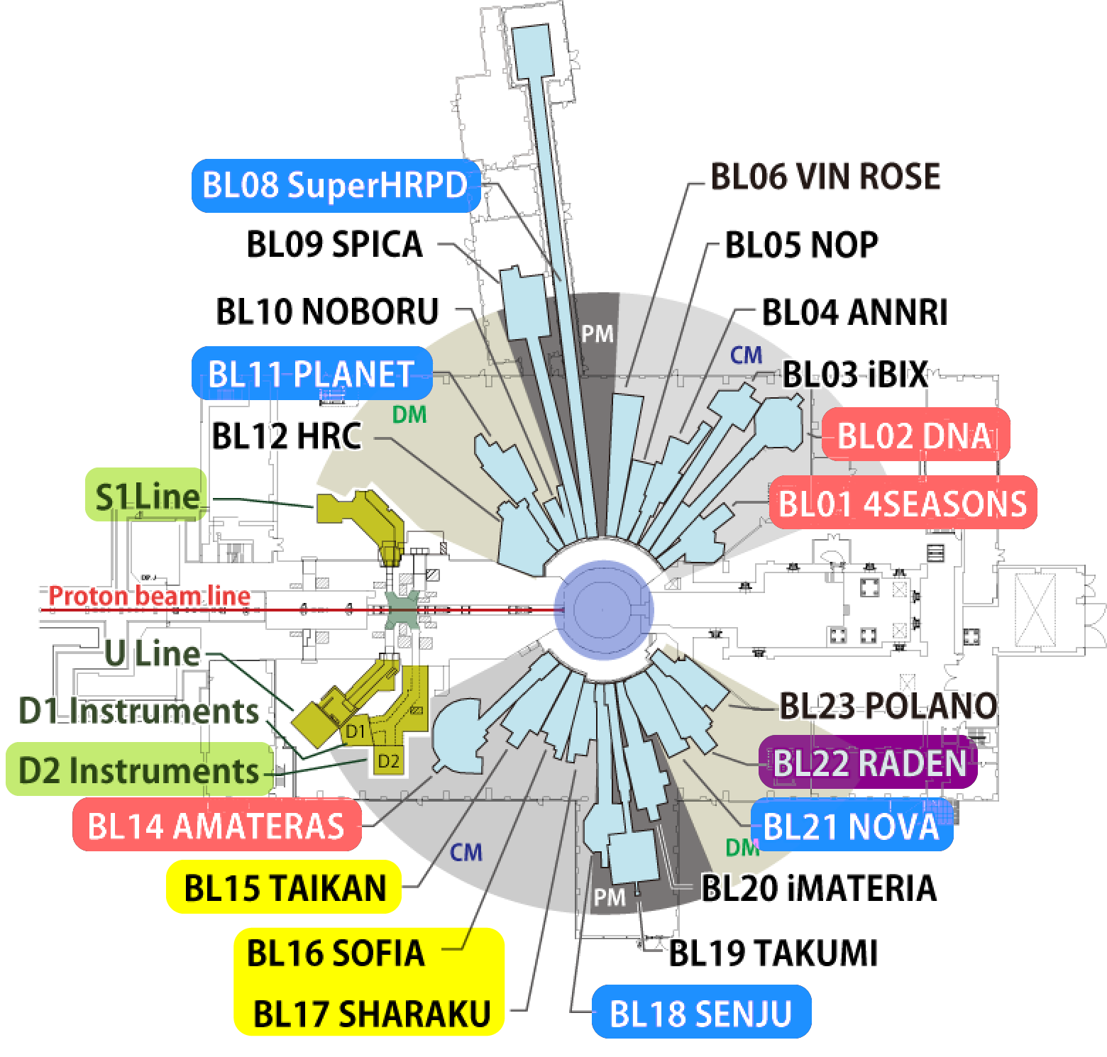
Course Descriptions
Inelastic Neutron Scattering
Diffraction
SANS/Reflectometry
Elemental analysis/Imaging
Muon

BL01: 4SEASONS
The Study of Lattice Dynamics in a Crystalline Material using Inelastic Neutron Scattering
Inelastic neutron scattering is an experimental method that is used to observe micro-vibration (dynamics) of atoms and spins in a material. By observing the difference in energy between the incident and scattered neutrons, the magnitudes, distances, and directions of the forces acting between the atoms or spins in the material can be determined.
In this course, students will learn how to measure atomic vibrations (phonons) in a single crystal as a function of momentum and energy by inelastic neutron scattering using the chopper spectrometer 4SEASONS. By analyzing the data, students will learn what kinds of forces act between atoms and how they affect the macroscopic properties.
BL02: DNA
The Study of Molecular Dynamics on the Nanosecond Timescale using Quasielastic Neutron Scattering
Quasielastic neutron scattering is considered to be one of the most effective techniques for measuring the dissipation motion (e.g. fluctuation or diffusion) of atoms, molecules and spins in a material. In particular, in a number of widely-used functional materials, such as in lithium secondary batteries or fuel cells, solid state ionic conductors play an important role. In these solid state ionic conductors, the ions or hydrogen atoms are moving at a speed similar to that of the liquid state, even at around room temperature. These dynamic motions of ions and hydrogen atoms can be measured at the nanosecond timescale using quasi-elastic neutron scattering.
In this course, students will study the hydrogen ion dynamics in a Nafion ion exchange membrane, a material that is currently in practical use in polymer electrolyte fuel cells, by the DNA high-resolution spectrometer. At the same time, students will learn how to analyze the data from a typical quasielastic neutron scattering experiment.
BL14: AMATERAS
Study of Spin Dynamics by using Inelastic Neutron Scattering
By using inelastic neutron scattering technique, we can observe motions of atoms and spins (atomic magnets) in a material. Neutron scattering data tell us how these atoms and spins are coupled with each other in the sample. We can extract lots of underlining microscopic information, which is indispensable to material science studies, from the data.
In this course, students will learn about how to measure the collective motions of spins in a crystalline sample on AMATERAS, a cold-neutron disk-chopper-type inelastic neutron scattering instrument. The students can experience basic of study on spin dynamics in a crystalline system through a simulated experiment on a simple spin system and data analysis.
BL08: SuperHRPD
Decipher the Materials Structure of Interest
We are surrounded by materials with various functions. Functions of batteries, magnets, superconductors, multiferroic materials, optical & thermoelectric materials, soft materials, etc. are consequences of atomic structures & their changes with various characteristic scales.
In this course, we will learn about the method to decipher the atomic structures of functional materials using advanced neutron diffraction techniques. Understanding atomic structures are the first step in materials science. Students will conduct high resolution neutron diffraction data analyses using both the SuperHRPD diffractometer (beamline BL08) and the SPICA diffractometer (BL09).
BL11: PLANET
In-situ Pressure Neutron Diffraction
Pressurization reduces interatomic distance of materials, which often induces drastic changes in their physical properties. To understand the origin of the changes, its structural information is essential. In-situ high-pressure neutron diffraction useful for getting the information.
In this course, participants will learn methods to generate pressure and determine crystal structures through analyses of the powder diffraction data of a high-pressure polymorph, ice VII.
BL18: SENJU
Structural analysis of single crystals using neutron diffraction
Physical properties and functionalities of materials are intrinsically linked to the arrangement of atoms inside the materials. Diffraction methods are frequently used to determine the atomic-scale structure of crystalline materials, and neutron single crystal diffraction is particularly well-suited to the study of materials where the functionality depends on the position of light elements and/or the arrangement of magnetic moments.
In this course, basic lectures about neutron single crystal structure analyses using BL18 (SENJU) will be given followed by actual data reduction and data analysis exercises.
BL21: NOVA
Local Structure Analysis of Disordered Materials by High Intensity Total Diffractometer NOVA
Neutron total scattering is a powerful tool for the structure analysis of disordered materials including amorphous and liquid.
The technique obtains a pair correlation function by Fourier transformation of the measured scattering cross-section from which it is possible to discuss real space disordered atomic arrangement or (magnetic) local lattice distortion in the material.
A neutron total scattering instrument (NOVA) realizes measurements of a high resolution pair correlation function. In this course, participants will learn how to obtain a static structure factor and a pair correlation function of various disordered materials.
BL15: TAIKAN
Structural Analysis using the Small and Wide Angle Neutron Scattering Instrument TAIKAN
The small-angle neutron scattering (SANS) method is valuable in characterizing the nanoscale structure of materials. The Small and Wide Angle Neutron Scattering Instrument TAIKAN can probe structures in a sample on a correlation length scale from about 0.1 nm to over about 1,000 nm.
The following topics will be covered in this course:
· SANS method using a pulsed beam and Data analysis procedures
· Similarities and differences between SANS using a pulsed beam, SANS using a continuous beam and SAXS
· Diversity of sample environments
BL16: SOFIA, BL17: SHARAKU
Surface and interface analysis using neutron reflectometry
As different materials meet at surfaces and interfaces, they show characteristic properties and various functions due to their peculiarity, which attract chemists, biologists, and physicists. Neutron reflectometry (NR) is a powerful tool for investigating the surface and interfaces of soft matters, magnetic materials etc. on the nanometer to sub-micrometer length scale with taking advantage of the unique characteristics of neutrons. Neutrons can distinguish an interesting part labeled with deuterium and/or can observe an interface between solid and liquid through a substrate. Polarized neutron reflectometry can observe magnetic moment behavior on the surfaces and interfaces of magnetic materials.
In this course, students will perform virtual experiments of the NR experiments with non-polarized and polarized neutrons using the SOFIA and SHARAKU reflectometers, respectively. For the SOFIA, the neutron reflectivity profiles of polymer thin films on Si substrates in air and in water with different contrasts will be analyzed. For the SHARAKU, the neutron reflectivity profiles of magnetic thin films will be analyzed. The Parratt formalism will be used to calculate the reflectivity profiles and determine the change in the structure of the thin films.
BL22: RADEN
Visualization of Structure and Physical Property Distributions Using Pulsesd Neutron Imaging
Neutron imaging is a widely used nondestructive inspection method to visualize internal structure of objects by utilizing the neutron's unique characteristics such as large transmission power for heavy elements and high sensitivity to light elements like hydrogen. In addition, an energy resolved neutron imaging method, which has been developing in recent years, enables us to visualize not only the structure but also the spatial distribution of crystallographic information (Bragg-edge imaging method), elemental and thermal information (Resonance absorption imaging method), and magnetic information (polarized neutron method).
In this course, you will learn the following contents; the principle of pulsed neutron imaging for both conventional and the energy-resolvedtechnique, the basis of neutron imaging experiment: the experimental setup, detector system and related devices, and data processing and visualization.
S1: ARTEMIS
Positive muon spin relaxation (μSR)
Positive muon in a material stops at an interstitial site, observes magnetic fields of the environment and exhibits Larmor spin precession. By measuring the decay positrons emitted from muons, time dependent behavior of the muon spin in a material is known. This is the spectroscopy called (positive) muon spin relaxation (μSR). This technique yields the information of the magnetic property of a material, including magnetism and superconductivity and the hydrogen state in a material with the muon being a light hydrogen isotope.
In contrast to neutron, muon is a local magnetic probe in real space with a unique time scale, being a powerful probe of spin relaxation phenomena.
In this course, students will perform simulated μSR measurement at the S1-ARTEMIS spectrometer and will receive instruction of data analysis. Introductory lectures on μSR and other muon measurements will also be given as a part of the school.
D2
Non-destructive elemental analysis using muonic X-ray
The negative muon is a lepton particle with a charge of -e, a spin of 1/2 and a mass of approximately 200 times larger than that of the electron. When a negative muon is irradiated into a matter, it replaces a bound electron and forms a bound state with a nucleus. Then, as in the case of electrons, the muon finally falls to the ground state with emitting characteristic X-rays attributed from negative muons (muonic X-ray). Elemental analysis can be performed using these muonic X-rays.
The feature is that the energy of muonic X-rays are 200 times larger than that of the characteristic X-rays derived from electrons (electron X-ray). The high-energy muonic X-rays can escape to the outside of a sample even if it is from the inside where the electron X-rays are absorbed. Therefore, the emitted muonic X-rays are detectable using Ge-detectors. That is why nondestructive analysis is possible even inside a sample that cannot be analyzed by electron X-rays.
In this course, participants will receive introductory lectures, including the features of elemental analysis using muonic X-rays and experimental method, and perform elemental analysis of unknown samples.
 The 4th